CT Users Group
CT Users Group meeting information
Home
CTUG home page
About
About the CTUG
Meetings
Info and talks from meetings
Contact
How to contact us
Mail list
ctusers mail list
Links
Some useful links
Search
Search and site map
Meetings » 14th CTUG Meeting » Abstracts
Optimisation of the Philips CT automatic exposure control system
T Wood
Hull & East Yorkshire Hospitals
Abstract
Introduction: The purpose of this study was to develop a technique for optimising the automatic exposure control (AEC) system on three Philips CT scanners (two Brilliance 16 and one Brilliance 40). This included matching the dose and imaging performance of the DoseRight Automatic Current Selection (ACS) system, and the longitudinal and rotational modulators (Z-DOM and D-DOM).
Method: The Rando anthropomorphic phantom was scanned using appropriate clinical settings to define the 'user determined image noise reference'; the ACS system uses this to determine the maximum mAs that is required to produce clinically acceptable images of real patients, based on the results of their topogram. This technique replaced the 'automatic patient size averaging' system that is the default option on these types of scanner, but was extremely difficult to optimise due to the way in which it is always evolving. The response of the system in terms of radiation dose and image noise was then assessed as a function of 'patient size' with a purpose built AEC phantom. The performance of each scanner was then compared to ensure performance was well matched across the clinically relevant range of phantom thickness, and adjustments made to the ACS settings where necessary.
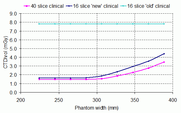
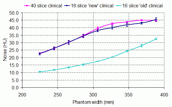
Results: Despite the complexities of the AEC implementation, introduced through the use of a 'user determined image noise reference' (as opposed to a single image quality metric, such as standard deviation), it was possible to match the performance of these systems through the use of simple phantoms (Figure 1). The use of the Rando anthropomorphic phantom allows easy adjustments to be made to the reference image for the purposes of optimisation (e.g. reducing mAs), as it does not suffer the significant inter-patient variations associated with the use of real clinical images. Several limitations of the AEC system have also been identified through a detailed analysis of the image noise properties.


Figure 1: Optimisation of the 'Chest only' protocol on a 16 slice Philips Brilliance CT scanner. Prior to setting up the ACS system this scanner used a fixed mAs technique (light blue line), but after using the Rando phantom to define the 'image noise reference' (dark blue line), performance is now well matched to a 40 slice system setup in the same way (magenta line). (a) A plot of radiation dose (CTDIvol) and (b) image noise (as defined by the standard deviation) as a function of phantom width.