28th CT users group meeting: 22/10/2026
The twenty-eighth CTUG meeting will be held in Leicester on Thursday 22nd Octover 2026 at St Martin’s House conference centre, 15 minute’s walk from Leicester railway station and close to parking in the centre of town (although please note that King Richard III’s car park is now a museum, just across the road from the venue)!
The draft programme is shown below, with abstracts available (press the +/- buttons!).
Online registration is now available for the 2026 annual meeting and CTUG Training Course. The cost for attendance at the CTUG annual meeting is £50, including refreshments and lunch.
The day before the meeting (21st October), we will run our third one-day training course - described as “An introduction to the physics of computed tomography including hardware, image formation, and factors affecting radiation dose and image quality”, check out the CTUG training course page for more details.
| « Previous meeting | – | Meetings Index | – | Next meeting » |
|---|
CT USERS GROUP - ANNUAL MEETING DRAFT PROGRAMME
09:30 Registration – Tea and Coffee
09:55 Welcome and introduction - Laurence King - Royal United Hospitals Bath NHS Foundation Trust
Session 1 - Evaluating CT performance
10:00 Evaluating whether SilverBeam can be used to reduce dose whilst maintaining image quality for a chest CT protocol - Kristina Timkovicova, Hannah Dickens, David Platten - United Lincolnshire Teaching Hospitals NHS Trust Purpose
To evaluate whether SilverBeam can be used to reduce dose whilst maintaining image quality for a low-dose chest CT protocol.
Methods
A CT dose index (CTDI) phantom was used to confirm the accuracy of the displayed volume CTDI (CTDIvol) by comparing it to a calculated value. A Gammex Mercury 4.0 phantom and a Kyoto Kagaku N-1 LUNGMAN chest phantom were scanned using a clinical CT chest protocol and a modified SilverBeam protocol. Tube current was fixed to achieve displayed CTDIvol values of 1.3-0.1 mGy when scanning with SilverBeam. A detectability index (d’) was calculated for each set of images of the Mercury phantom. d’ when using SilverBeam was compared to d’ when using the clinical protocol. Images of the chest phantom, with and without a breast plate, were scored based on image quality and artefacts. The number of visible simulated tumours were also noted.
Results
The measured CTDIvol was 0.4% higher than the displayed value without SilverBeam and 5.2% higher with SilverBeam. d’ values at each dose level with SilverBeam were all statistically significantly lower than the d’ result of the clinical protocol (p < 0.0001). The 1.1 mGy images of the chest phantom without the breast plate received the highest image quality score (3.7/4). SilverBeam reduced streak artefacts through the shoulders in the 1.3 mGy and 1.1 mGy images. Most scorers correctly located all simulated tumours in all sets of images.
Conclusion
d’ with SilverBeam was not comparable to the clinical protocol. The chest phantom images suggested that the difference in image quality may not be clinically significant.10:20 Matching reconstruction kernels based on noise power spectrum - Platten DJ, Smith E, Dickens H - United Lincolnshire Teaching Hospitals NHS Trust Introduction
Canon have introduced a new CT scanner software platform called INSTINX [1]. Canon have not provided a mapping between the anatomical kernel names on the INSTINX platform and the kernels on the previous platform. This mapping is required to harmonise scan protocols between INSTINX and non-INSTINX scanners.
This work has used the measured noise power spectrum (NPS) to match each anatomically named INSTINX reconstruction kernel to an FC kernel.
Method
A Phantom Laboratory Catphan 500 phantom was scanned on Canon Aquilion Serve SP (INSTINX) and Canon Aquilion Prime SP (non-INSTINX) CT scanners using a standard acquisition protocol. Scans were repeated for each available reconstruction kernel (n=20 Serve SP; n=55 Prime SP).
For each kernel the NPS was calculated from images of the uniform section of the Catphan using an ImageJ plugin run in batch mode [2, 3].
For each Serve SP kernel NPS the closest matching Prime SP kernel NPS was found using a score that was a combination of root mean square error and Pearson correlation coefficient [3], a similar but more simplistic approach to that of Solomon et al [4]. Plots of all pairs of Serve SP and Prime SP NPS results were produced to enable a visual check of the numerical scoring.
Results
The top scoring kernel was a good visual match for 19 of the 20 Serve SP protocols. For the remaining Serve SP kernel the highest scoring Prime SP kernel visual match was poor. For this kernel the best visual match was the 4th highest score.
The method presented has provided a robust way of matching CT reconstruction protocols between two different models of Canon CT scanner based on NPS measurements. The method may also be useful to find best matches between scanners from different vendors. DICOM images, analysis tools, and results of this work are available on Bitbucket [2, 3].
References
1. INSTINX, https://eu.medical.canon/innovations/efficient-workflow/, accessed 13/7/2026
2. ImageJ NPS plugin, https://bitbucket.org/dplatten/imagej-plugins/
3. ImageJ Jython NPS batch processing plugin and NPS matching Python script, https://bitbucket.org/dplatten/ct-nps-matching/
4. Solomon JB, Christianson O, Samei E. Quantitative comparison of noise texture across CT scanners from different manufacturers. Medical Physics. 2012 Oct;39(10):6048–55. https://doi.org/10.1118/1.475220910:40 The development of a local testing methodology for Automatic Tube Current Modulation (ATCM) in Computed Tomography (CT) - Tobias Woodford, Anne Hill, Anna Darlington - University Hospitals Bristol & Weston NHS Foundation Trust Aim
To identify and evaluate the most suitable phantom and testing methodology for characterising the performance of ATCM systems used in CT.
Background
As the frequency of CT examinations continues to rise, and given the relatively high radiation doses involved, technical solutions for dose optimisation have become increasingly important to mitigate excessive radiation exposures to patents; one such technical solution is ATCM. Currently there is no specific UK guidance on how to reliably and consistently test ATCM; however, ensuring a robust testing methodology using an appropriate phantom is essential and should form part of a routine CT quality assurance testing programme. An appropriate phantom should simulate the changes in attenuation along its length that would be observed in a patient examination.
Methods
Three phantoms were assessed for suitability; a CTDI phantom (Gammex, USA), a Mercury phantom (Sun Nuclear Corporation, USA) and a CelT phantom (Betsi Cadwaladr University Health Board, UK). The phantoms were scanned on a Canon Aquilion One. Each phantom was scanned with varying parameters to identify the most appropriate factors for routine ATCM testing, after which a consistent set of optimised exposure settings were applied to each phantom, enabling a fair comparative assessment of their performance. As a guide on characterising ATCM, the American Association of Physicists in Medicine (AAPM) report 233 was used. The phantoms were scanned several times with identical settings to assess repeatability of results.
Analysis of 65 datasets were completed using ImageJ (National Institutes of Health, USA) software. Regions of interest (ROI) were taken on each slice throughout the scan volumes. For the CTDI phantom, two large circular annular ROIs were drawn. For the Mercury and CelT phantoms, four small ROIs were drawn in the cardinal positions within every image slice, with care taken to avoid including the inserts of the test object within the ROIs. The Hounsfield Unit and standard deviation were evaluated for each ROI. The mA was extracted from the DICOM header. ATCM performance was assessed by plotting these three parameters against slice location. The dependence of tube current and image noise on water equivalent diameter will be investigated.
Results
All three phantoms successfully demonstrated ATCM performance, with tube current varying in response to changes in attenuation. The Mercury phantom provided the most comprehensive technical assessment and was suitable for statistical analysis of the relationship between ln(mA) and water-equivalent diameter. The CelT phantom demonstrated good repeatability and provided the clearest demonstration of ATCM behaviour, whilst also being the easiest to set up and position. The CTDI phantom produced repeatable results but its abrupt changes in attenuation limited its ability to characterise ATCM performance consistently across the entire scan length.
Discussion
This project will discuss the suitability of different phantom designs for routine ATCM testing, with emphasis on the practicality, measurement repeatability, and the clinical relevance of the phantoms; highlighting the benefits and limitations of each. The discussion will explore the relationship between image noise and tube current plotted against water equivalent diameter, in line with the analysis outlined in AAPM Report 233.
Conclusion
All three phantoms were suitable for assessing ATCM performance. The Mercury phantom provided the most comprehensive technical assessment; however, when both technical performance and practical usability were considered, the CelT phantom provided the best balance and was considered the most suitable for routine ATCM quality assurance testing. The CTDI phantom remains useful but is less suitable for detailed ATCM characterisation due to its discrete attenuation structure.
With special thanks to Royal United Hospitals Bath NHS Foundation Trust and Velindre University NHS Trust for enabling this project through the loan of their phantoms.
Key references
[1] AAPM Reports - Performance Evaluation of Computed Tomography Systems - The Report of AAPM Task Group 233 https://www.aapm.org/pubs/reports/RPT_233.pdf
[2] Merzan, D, Nowik P, Poludniowski G, Bujila R. ‘Evaluating the impact of scan settings on automatic tube current modulation in CT using a novel phantom’, Br J Radiol 2017: 90: 20160308 https://doi.org/10.1259/bjr.2016030811:00 Tea and coffee
Session 2 - Advancing CT Technologies
11:30 Experience with Siemens Photon Counting CT - Alexandra Palmer - Barts Health NHS Trust Abstract to follow
11:50 Optimisation of reconstruction matrix size in photon counting coronary CT angiography - Anna Wang, Jamie Dormand, Md Imran Hossain, Jonathan Weir-McCall - The Royal Marsden NHS Foundation Trust Coronary CT angiography (CCTA) is recommended for assessing coronary artery disease, but the spatial resolution of conventional CT limits evaluation of stents to diameters > 3 mm [1]. Photon-Counting CT (PCCT) offers improved spatial resolution and matrix sizes up to 1024 are now available to capture these finer details. However, to fully realise these benefits, reconstruction parameters such as matrix size must be optimised to avoid unnecessary increases in data storage requirements.
Following initial clinical feedback that matrices larger than the vendor default improved image quality, we evaluated reconstruction matrix size in PCCT-CCTA using phantom and clinical datasets. PCCT-CCTA scans of an anthropomorphic chest phantom and Mercury 4.0 phantom were acquired using standard and ultra high-resolution (UHR) protocols [2]. Suitable clinical datasets were identified by a cardiologist and saved on the scanner for subsequent retrospective reconstruction. Images were reconstructed using 512, 768 and 1024 matrices across kernels Bv56-84. Modulation transfer function and noise power spectrum measurements of the iodine insert within the Mercury phantom were performed to compare reconstructions. Two experienced readers assessed the overall image quality, sharpness and noise for chest phantom and retrospectively reconstructed clinical datasets. Optimal matrix size was determined by comparing evaluations for image sets differing only in reconstruction matrix.
Results demonstrated that a 768 matrix provided the best balance between spatial resolution and image storage capacity in both UHR and standard CCTA datasets. Increasing the matrix size further to 1024 offered no meaningful improvements to image quality but approximately doubled storage requirements. Image noise was found to vary minimally between matrices. The 768 matrix differs from the vendor default and has since been implemented clinically with positive initial feedback.
References
[1] C. Vrints et al., ‘2024 ESC Guidelines for the management of chronic coronary syndromes: Developed by the task force for the management of chronic coronary syndromes of the European Society of Cardiology (ESC) Endorsed by the European Association for Cardio-Thoracic Surgery (EACTS)’, Eur. Heart J., vol. 45, no. 36, pp. 3415–3537, Sep. 2024, https://doi.org/10.1093/eurheartj/ehae177
[2] ‘Mercury 4.0 Phantom (GammexTM Technology) - Sun Nuclear’. Accessed: Aug. 19, 2025. [Online]. Available: https://www.sunnuclear.com/products/mercury-4.0-phantom12:10 Commissioning photon-counting CT: Lessons from our experience in driving clinical engagement - Ruby Callister, Louise Giansante, Anna Wang, Jamie Dormand, Ed McDonagh - The Royal Marsden NHS Foundation Trust Photon-counting CT (PCCT) represents a major technological advancement in CT, offering improved spatial resolution, intrinsic spectral imaging, and potential for lower radiation doses. However, these benefits come at a substantial cost compared with conventional energy-integrating detector CT systems. Previous experience in the UK has also demonstrated that advanced CT capabilities, most notably dual-energy CT, can remain significantly underutilised despite clear clinical advantages. There is therefore a risk that, without deliberate implementation strategies, PCCT may not achieve its full clinical potential. Maximising value from this investment requires not only robust technical commissioning, but also active facilitation of clinical engagement and adoption.
This work describes our experience commissioning clinical PCCT systems across an oncology hospital and a specialist heart and lung hospital. Whilst the advantages of PCCT are well evidenced in cardiac imaging, their translation into oncology is less so. Consequently, the clinical value achievable with PCCT was highly dependent on our approach to implementation. Recognising this, we took up an active role in driving multidisciplinary engagement.
We will share successes, challenges encountered, areas for improvement and where luck was on our side. We will reflect on our role as medical physicists in facilitating this process, including the time investment required and the longer-term clinical benefits achieved. Using our experience, we aim to give others practical ideas for how medical physicists can add tangible value in translating novel CT technologies into routine clinical practice.12:30 Lunch
13:30 AGM and officer election
Session 3 - Optimisation and patient dose
14:00 Dose implications for incorrect protocol selection for paediatric head CT - Heloise Carpenter - Barking, Havering and Redbridge University Hospital Trust From completing a Barking, Havering and Redbridge University Hospital Trust (BHRUT) paediatric CT head dose audit, it became apparent that only 52% of examinations were completed on a paediatric protocol thus leading to large variation in radiation doses due to incorrect protocol selection. The audit included all under one year old paediatric patients receiving head CTs over the period of 15 months and included 65 patients. The variation in the Dose Length Products of these examinations in the audit were between 92 mGycm and 1016 mGycm. The use of adult protocols included using the default adult settings and adjusted protocols for a smaller patient size.
The dose implications of incorrect protocol selection were investigated by measuring the dose with the RaySafe X2 CT sensor for different protocols using a paediatric CTDI phantom on a Canon Aquilion One CT scanner and a Kyoto Kagaku Newborn Whole Body Phantom on a GE Revolution Ascend scanner. The results showed that the paediatric protocols using a static tube current were always the lowest dose protocol and that changing the default settings on the adult protocol using tube current modulation always resulted in a higher radiation dose. For the Canon Aquilion One CT, there was a 50% dose increase from selecting an adult protocol in comparison to the paediatric protocol whilst for the GE Revolution Ascend, this resulted in an 139% increase.
Dose calculations were also completed (using ImPACT and conversion factors) for the 65 patients showing a variation in effective doses from 0.67 mSv to 8.41 mSv (using ImPACT) and 3.6 mSv to 39.6 mSv using paediatric conversion factors published in by P.Chu et al in 2023. The average difference in dose between the two methods was a factor of 6. To further investigate differences in paediatric effective dose calculations for CT head examinations using Virtual Dose software for 15 under one year old Paediatric CTs were compared to calculated doses from ImPACT and conversion factors. Due to long latency periods of radiation induced cancer, patients in these cohort have an increased risk due to their expected life expectancy and due to their difference in size to adult patients, incorrect protocol selection impacts these patients more significantly than older paediatric patients.14:20 Experience in developing imaging protocols for pregnant patients in PET/CT - J. Edwards[1], D Sharkey[1,3], J Cole[1], B Ferreira[2], B Holman[2], T Willson[2], T Wagner[2] - [1]Radiological Physics, Royal Free London, [2]Department of Nuclear Medicine, Royal Free London, [3]Guys and St Thomas’ Hospital Introduction
The use of CT to image pregnant patients has typically been reserved for emergency or trauma indications due to the potential for high foetal exposures. The use of PET/CT in pregnancy is also generally avoided for similar reasons. Advances in technology, from both the CT and the PET perspective, mean imaging pregnant patients for specific clinical indications is becoming a more viable option. Since 2024 Royal Free London has been a referral centre for pregnant patients requiring PET/CT scans for Oncology imaging. The main aim of this work is to optimise CT parameters for imaging pregnant patients while providing adequate image quality for image interpretation.
Method
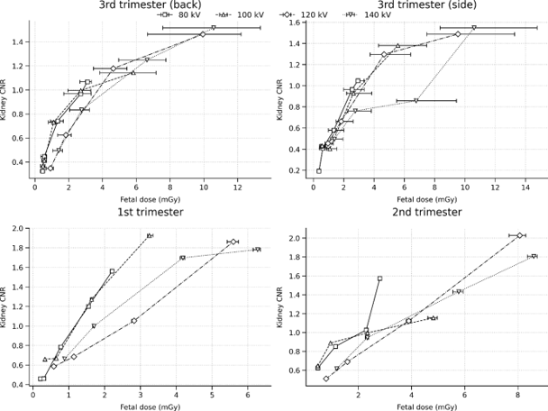
A whole-body and neonatal anthropomorphic phantom were used to simulate different stages of pregnancy. Scans were performed for simulated 1st, 2nd and 3rd trimester pregnancy at different dose levels. Image quality metrics were analysed and foetal doses estimated for each of the scans performed. This was used to provide feedback to the Nuclear Medicine clinicians to help inform future scanning protocols.
Results and Conclusions
For certain clinical indications the use of PET/CT in pregnancy can be achieved at significantly reduced doses compared to standard PET/CT imaging. Initial results suggest higher patient and foetal doses are required to maintain CNR in late-stage pregnancy compared to first and second trimester models. There is also some suggestion that CNR can start to decline for higher foetal doses.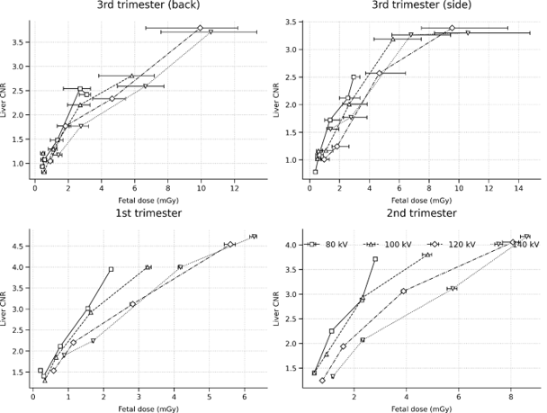
Although some impact to the CT image quality is observed, overall outcomes with fused images are acceptable from the clinician’s perspective. Foetal doses have not been shown to be significant and are generally below 1mGy. This work has also been used in the development of the National Total Body PET/CT Pregnant Patient Imaging Network which will also be discussed.14:40 NDRLs: Racing to the bottom while missing half the picture - John Loveland - Maidstone and Tunbridge Wells NHS Trust Abstract to follow
15:00 Tea and coffee
Session 4 - Lung screening CT
15:30 National survey of CT lung cancer screening scan protocols and participant doses - Gareth Iball, Anna Sharman - University of Bradford Background
The NHS England national lung cancer screening programme (LCSP) has now been rolled out across more than 50% of England and the expectation is that 100% roll out is achieved by 2030. Within Scotland, Wales and Northern Ireland, roll out of CT based lung cancer screening is planned or in the early stages of implementation. Participants in the LCSP are non-symptomatic, and as such there are particular concerns regarding radiation dose and risk, and these must be very carefully balanced with the need for high-quality images to enable confident diagnoses.
The current NHS England guidance on scan protocols, expected doses and image quality is open to misinterpretation and has led to variations in CT scanning practice across the programme. Similarly, evidence from a recent European survey showed notable variations in LCS CT scan protocols. Such variation has the potential to lead to inappropriate participant doses and or inadequate image quality which can limit the effectiveness of the imaging with the programme. To gain understanding of the current state of practice, NHS England LCSP has launched a national survey of CT scan protocols that are used for LCS and the resulting participant doses.
Method
A survey template was designed by the authors and NHS England LCSP staff. Data requested included specific details of the scan and reconstruction parameters for all LCS scan protocols, alongside the intended purpose of each reconstructed image set. The resulting CTDIvol, DLP & participant weight was requested for 100 participants on each scanner, regardless of participant weight.
The survey was distributed to all regional LCS programmes by the NHS England team on 6th July, with data to be returned by 14th August.
Results
A summary of the scan protocols will be presented, identifying variations in practice within individual programmes, and across similar scanner types that are used within the whole programme.
Participant dose data analysis will again explore variations between programmes and similar scanners. Dose changes as a function of participant weight will be explored and where possible doses will be compared with existing standards and evidence from the literature.15:50 Lung screening roundtable discussion
16:20 Close of meeting